Center for Augmented Intelligence in Imaging
Mayo Clinic Florida
Integrating and Deploying AI Models within Clinical-Imaging Workflows
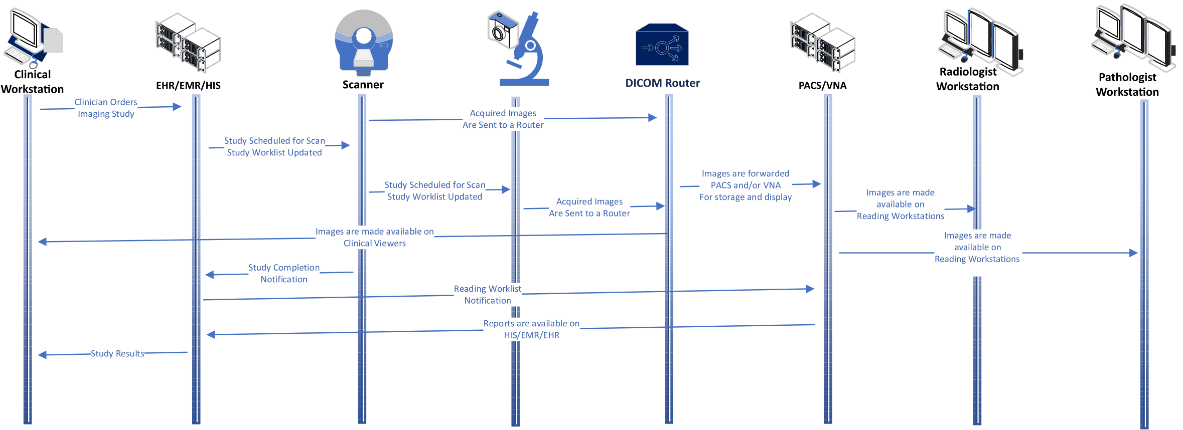
Effective integration of imaging-related (pixel- and nonpixel-based) Artificial Intelligence (AI) models into existing clinical Radiology workflows is critical since such additions can greatly impact (either positively or negatively) operational efficiencies or downstream decision making (e.g., surgery, pathology, interventions, and drug precautions) [1]. In order to facilitate seamless integration of imaging-AI capabilities, with minimal negative influence on existing Radiology workflows (Figure 1), the Center for Augmented Intelligence in Imaging (CAII) at Mayo Clinic Florida has developed infrastructure and modular software packages functionally compatible with MONAI [2] software packages (e.g., "MONAI Core" and "MONAI Deploy").
AI-based infrastructure should be both indistinguishable from the existing IT environment and require, at most, minimal training of Radiology users (e.g., radiologists and technologists). Nevertheless, the introduction of such tools requires the fostering of trust among the users as well as beneficiaries (e.g., patients and referring clinicians).
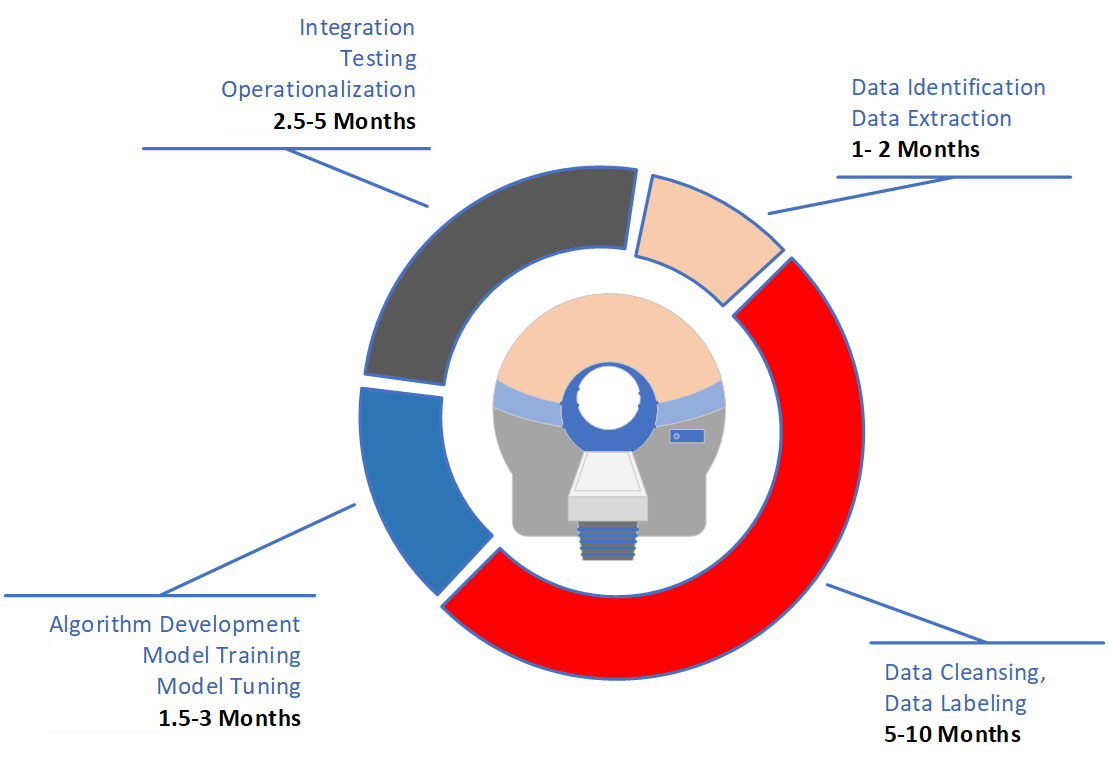
As the leading discipline in utilizing AI in medicine, Radiology has already recognized the need for greater efficiencies in all aspects of imaging-AI application, including AI-model development, deployment, and adaptation to real-world encounters. Unfortunately, these processes remain prohibitively time-consuming, laborious, and costly, often resulting in significant limitations to meaningful imaging-AI use (Figure 2).
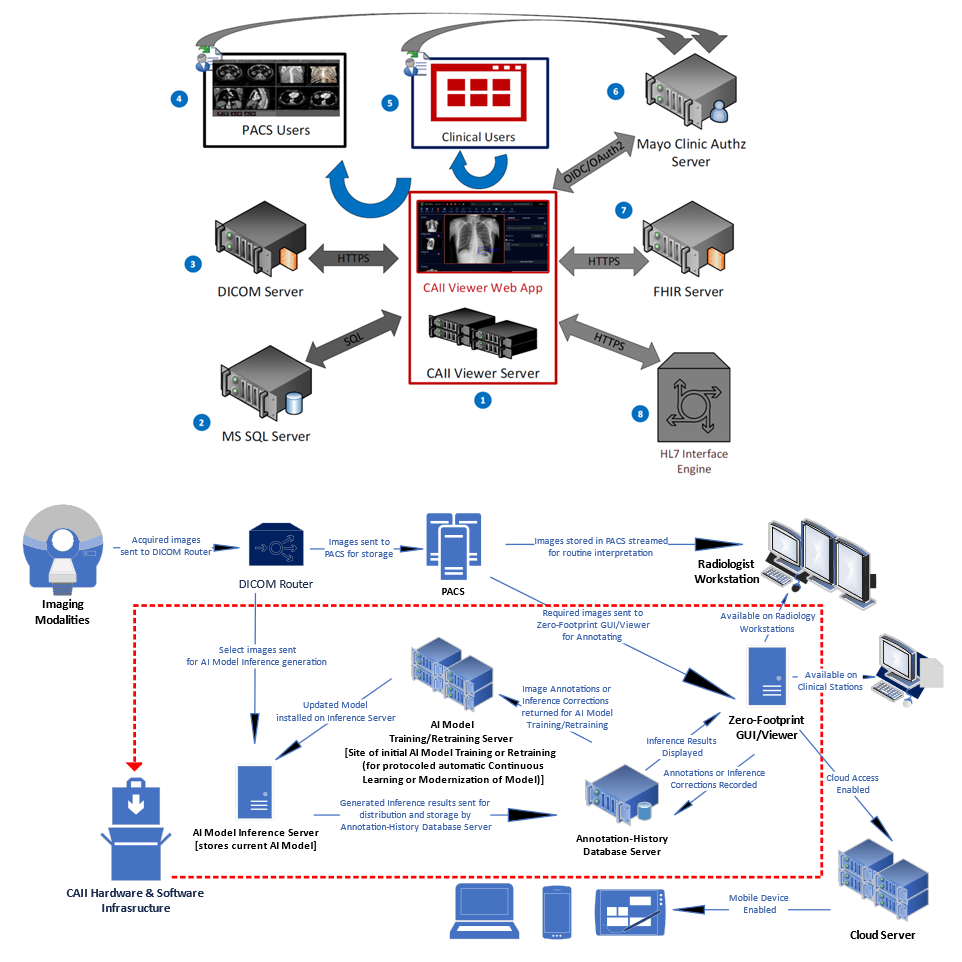
Engineers, imaging scientists, and physicians working in the CAII have developed infrastructure and containerized software packages, enabling imaging-AI models to be seamlessly integrated into the existing IT environment of a busy Department of Radiology [3-9]. The necessary interfaces and packages can be deployed on-premise, in-cloud, or in hybrid settings (Figure 4). The goal is to require minimal user training and IT support while fostering confidence in users and beneficiaries.
CAII at Mayo Clinic Florida has developed various capabilities to streamline the integration of imaging AI models into Radiology workflows. These capabilities include:
- Critical-results alerting
- Expert-in-the-loop AI-model deployment
- On-demand model training in clinical settings
- Real-time user inference-results adjudication with feedback in clinical settings
- Monitoring of user satisfaction
- Data collection for FDA approvals
- Continuous Learning
- Federated Learning
- Standards-based communication (DICOM, FHIR, HL7, IHE) between clinical systems
- Standards-based data collection regarding system and model performances
References
- 1. Gupta V, Erdal BS, Ramirez C, Floca R, Jackson L, Genereaux B, Bryson S et al. "Current State of Community-Driven Radiological AI Deployment in Medical Imaging." arXiv preprint arXiv:2212.14177 (2022).
- 2. Cardoso J, Li W, Brown R, Ma N, Kerfoot E, Wang Y, Murrey B et al. "MONAI: An open-source framework for deep learning in healthcare." arXiv preprint arXiv:2211.02701 (2022).
- 3. Testagrose C, Gupta V, Erdal BS, White RD, Maxwell RW, Liu X, Kahanda I, Elfayoumy S, Klostermeyer W, Demirer M. "Impact of Concatenation of Digital Craniocaudal Mammography Images on a Deep-Learning Breast-Density Classifier Using Inception-V3 and ViT." In 2022 IEEE International Conference on Bioinformatics and Biomedicine (BIBM), pp. 3399-3406. IEEE, 2022.
- 4. White RD, Demirer M, Gupta V, Sebro RA, Kusumoto FM, Erdal BS. "Pre-deployment assessment of an AI model to assist radiologists in chest X-ray detection and identification of lead-less implanted electronic devices for pre-MRI safety screening: realized implementation needs and proposed operational solutions." Journal of Medical Imaging 9, no. 5 (2022): 054504.
- 5. Gupta V, Demirer M, Maxwell RW, White RD, Erdal BS. "A multi-reconstruction study of breast density estimation using Deep Learning." arXiv preprint arXiv:2202.08238 (2022).
- 6. Demirer M, White RD, Gupta V, Sebro RA, Erdal BS. "Cascading neural network methodology for artificial intelligence-assisted radiographic detection and classification of lead-less implanted electronic devices within the chest." arXiv preprint arXiv:2108.11954 (2021).
- 7. White RD, Erdal BS, Demirer M, Gupta V, Bigelow MT, Dikici E, Candemir S, Galizia MS, Carpenter JL, O'Donnell TP, Halabi AH, Prevedello LM. Artificial Intelligence to Assist in Exclusion of Coronary Atherosclerosis During CCTA Evaluation of Chest Pain in the Emergency Department: Preparing an Application for Real-world Use. J Digit Imaging. 2021 Jun;34(3):554-571. doi: 10.1007/s10278-021-00441-6. Epub 2021 Mar 31. PMID: 33791909; PMCID: PMC8329136.
- 8. Rockenbach MABC, Buch V, Gupta V, Kotecha GK, Laur O, Erdal BS, Yang D, Xu D, Ghoshajra BB, Flores MG, Dayan I, Roth H, White RD. Automatic detection of decreased ejection fraction and left ventricular hypertrophy on 4D cardiac CTA: Use of artificial intelligence with transfer learning to facilitate multi-site operations. ntelligence-Based Medicine. 2022; 6.
- 9. Gupta V, Taylor C, Bonnet S, Prevedello LM, Hawley J, White RD, Flores MG, Erdal BS. Deep Learning Based Automatic Detection of Adequately Positioned Mammograms. Lecture Notes in Computer Science: Domain Adaptation and Representation Transfer, and Affordable Healthcare and AI for Resource Diverse Global Health. 2021; 12968:239-250.
Talks
Interested in Deploying Your AI Research?
Learn more about how MONAI Deploy can help scale your medical imaging AI applications across institutions worldwide.